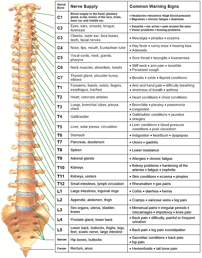
Anatomical Chart Spinal Nerves Spinal Nerve Root Innervation Chart Images Biology Diagrams Spinal nerve, in vertebrates, any one of many paired peripheral nerves that arise from the spinal cord. In humans there are 31 pairs: 8 cervical, 12 thoracic, 5 lumbar, 5 sacral, and 1 coccygeal. Each pair connects the spinal cord with a specific region of the body. Anatomy and Physiology - Spinal and Cranial Nerves; Ask the Chatbot a Question Several clinical considerations apply to spinal nerve anatomy. As previously mentioned, nerves exiting the cervical spine course horizontally from the spinal cord, exiting the intervertebral foramen. On the other hand, since the spinal cord usually extends caudally to the level of the L1 or L2 vertebral levels, the more caudal nerve roots
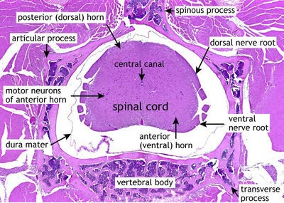
Spinal Nerve Branching. As shown in Figure \(\PageIndex{2}\), axons coming from the posterior (dorsal) root ganglion enter the posterior side through the posterior (dorsal) nerve root.The axons emerging from the anterior side do so through the anterior (ventral) nerve root.The posterior and anterior nerve roots fuse together to form the spinal nerves.

What They Are and What They Do Biology Diagrams
Nerves in Spine Anatomy. The spinal cord serves as a communication highway, linking the brain to the rest of the body through a network of nerve fibers. These fibers branch into pairs of nerve roots that exit through small openings (foramina) between the vertebrae. Each segment of the spinal cord connects to specific regions of the body, which

Anatomy of Spinal Nerves. Spinal nerves are large nerves that are distributed evenly along the spinal cord and the spine. The spine is a column of vertebrae bones that protects the spinal cord. These spinal nerves are large as they are formed by both sensory and motor nerve roots merging together. These nerve roots emerge from the spinal cord

Definition, Function, Diagram, Number, & Facts - Britannica Biology Diagrams
For the most part, the spinal nerves exit the vertebral canal through the intervertebral foramen below their corresponding vertebra. Therefore, there are 12 pairs of thoracic spinal nerves, 5 pairs of lumbar spinal nerves, 5 pairs of sacral spinal nerves, and a coccygeal nerve. The cervical spinal nerves differ from this pattern. C1-C7 spinal A spinal nerve is a mixed nerve, which carries motor, sensory, and autonomic signals between the spinal cord and the body. In the human body there are 31 pairs of spinal nerves, one on each side of the vertebral column. [1] [2] These are grouped into the corresponding cervical, thoracic, lumbar, sacral and coccygeal regions of the spine. [1]There are eight pairs of cervical nerves, twelve