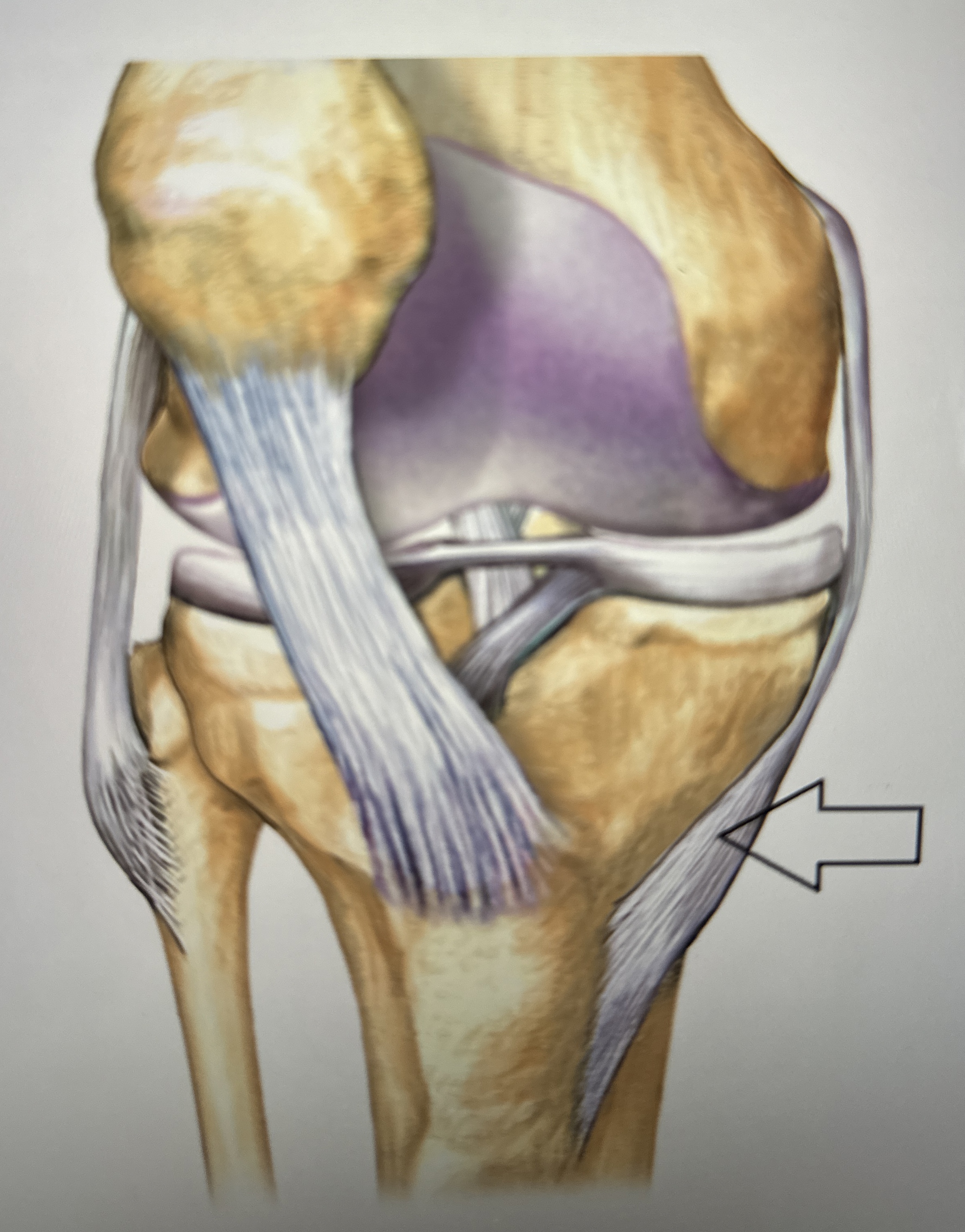
Your muscle attachment points influence your strength Biology Diagrams Unlike other ligaments or tendons, the anterior cruciate ligament normally has a heterogeneous appearance and the anteromedial and posterolateral bundles are defined by surrounding high-intensity structures 1.. The ACL Blumensaat line angle is normally ≤15º. It is calculated by drawing a line parallel to the roof of the intercondylar notch of the femur (Blumensaat line) and one parallel to

The inguinal ligament or Poupart's ligament formed from the aponeurosis of the lower border of external obliquis muscle. It has 2 surfaces concave and convex, the convex surface toward the thigh attached to the deep fascia that pulling the ligament downward. It is a site for attachment of internal obliquis muscle at the lateral 2/3 and transversus abdominis at the lateral 1/3. It is caused by axial traction or a sudden pull of the extended pronated arm. The radial head moves out of the weak annular ligament and capitellum, resulting in slipping over and subluxation of the radial head into the supinator muscle and annular ligament. Assessment [edit | edit source] Typical history might include a sharp jerk to the arm.
Radiology Reference Article - Radiopaedia.org Biology Diagrams
The inguinal ligament is involved in the condition known as the sportsman groin (inguinal disruption). Additionally, this ligament is an important surgical landmark and a groin hernia repair component. About 20 million people have an inguinal hernia repair yearly, making this type of surgery one of the most commonly performed worldwide. The lifetime occurrence of groin hernia, including These are the attachment points for tendons and ligaments. In general, their size and shape is an indication of the forces exerted through the attachment to the bone. A hole is an opening or groove in the bone that allows blood vessels and nerves to enter the bone. As with the other markings, their size and shape reflect the size of the vessels

Inguinal ligament (Ligamentum inguinale) The inguinal ligament (also ligamentum inguinale, arcus inguinalis or Pouparts's ligament) is a band of connective tissue that extends from the anterior superior iliac spine of the ilium to the pubic tubercle on the pubic bone.. It is formed by the free inferior border of the aponeurosis of the external oblique muscle which attaches to these two points. Its attachment points stabilize the lower abdomen and pelvis, which is crucial for various activities and movements. Anterior Superior Iliac Spine to Pubic Tubercle. The inguinal ligament spans from the anterior superior iliac spine (ASIS), located on the pelvic bone's anterior aspect, to the pubic tubercle, a bony prominence on the pubic bone. The anterior cruciate ligament (ACL) is a band of dense connective tissue which courses from the femur to the tibia. It consists of type I Both the medial and lateral intercondylar tubercles of the tibia serve as the attachment points for the two anteromedial and posterolateral bundles of the ACL.. For more detail on the anatomy of the
